[OCT Observations] OCT-A and Diabetic Retinopathy
![[OCT Observations] OCT-A and Diabetic Retinopathy Image](https://blog.visionix.com/hs-fs/hubfs/Trend%20analysis%201-1.png?width=1250&name=Trend%20analysis%201-1.png)
Patient description
A 60-year-old woman presented for a follow-up consultation.
She is diagnosed Type 2 Diabetic Retinopathy with an history of PRP (panretinal photocoagulation) treatment. Her last HbA1C measured 7%. She had ongoing anti-VEGF therapy.
VA : 10/10 in 2016 VA : 8/10 in 2021
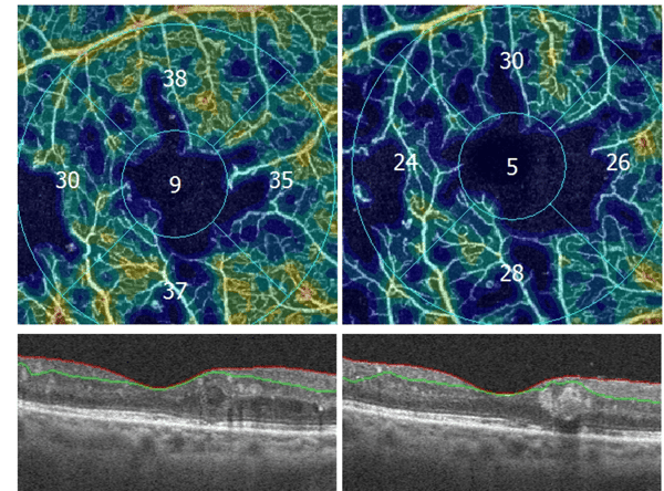
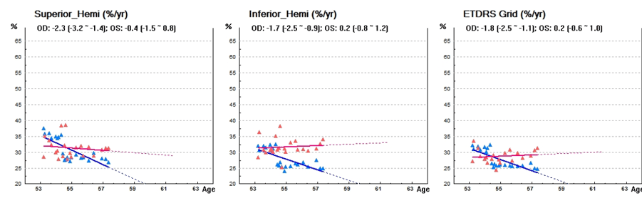
- Trend Analysis : Superficial capillary plexus vessel density – OD (1)
![]()
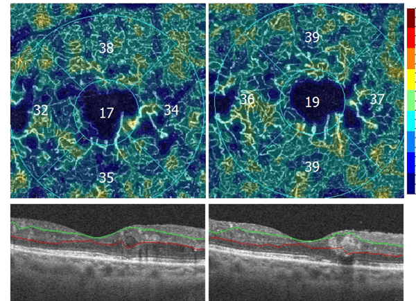
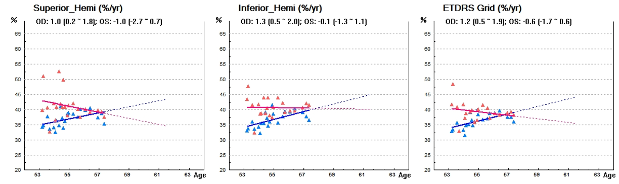
- Trend Analysis : Deep capillary plexus vessel density – OD (2)
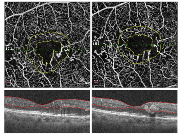
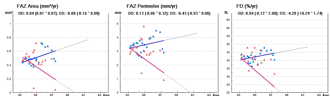
- Trend Analysis : Foveal Avascular Zone (FAZ) – OD (3)
- OCT-A dynamic 4 years follow-up (4):
Clinical Findings
- OCTA 4 years follow up of Diabetic Retinopathy type 1 shows capillaries drop out within superficial plexus with Vessel Density decreasing during anti-VEGF therapy (1). FAZ is enlarged and more prounonced 4 years after (3).
- Dynamic approach highlights vascular remodeling around foveal avascular zone in deep plexus (4).
- Vessel density in deep plexus is more stable and closes related to the conservation of visual acuity (2).
- Trend analysis confirms morphologic approach with superficial plexus decreasing and deep plexus stability.
Solix brings very high definition of superficial and deep plexus with the vessel density quantification. Trend analysis of each plexi confirms changes in each plexus and the correlation with visual acuity. Vascular drop out are clearly visible. SSADA algorithm brings more contrast to highlight the vascular remodeling during the follow up.
Conclusion
- Trend analysis is mandatory to confirm relevant changes within vascular plexi.
- Capillaries drop out occurs mainly in superficial plexus in diabetic retinopathy and during follow up this plexus is more affected.
- Preservation of deep plexus density in DR is correlated to a conservation of visual acuity
- OCTA is a good biomarker of DR progression
References
- Agemy SA, Scripsema NK, Shah CM, Chui T, Garcia PM, Lee JG, Gentile RC, Hsiao YS, Zhou Q, Ko T, Rosen RB. RETINAL VASCULAR PERFUSION DENSITY MAPPING USING OPTICAL COHERENCE TOMOGRAPHY ANGIOGRAPHY IN NORMALS AND DIABETIC RETINOPATHY PATIENTS. Retina. 2015 Nov;35(11):2353-63. doi: 10.1097/IAE.0000000000000862. PMID: 26465617.
- Mané V, Dupas B, Gaudric A, Bonnin S, Pedinielli A, Bousquet E, Erginay A, Tadayoni R, Couturier A. CORRELATION BETWEEN CYSTOID SPACES IN CHRONIC DIABETIC MACULAR EDEMA AND CAPILLARY NONPERFUSION DETECTED BY OPTICAL COHERENCE TOMOGRAPHY ANGIOGRAPHY. Retina. 2016 Dec;36 Suppl 1:S102-S110. doi: 10.1097/IAE.0000000000001289. PMID: 28005668.
- Dupas B, Minvielle W, Bonnin S, Couturier A, Erginay A, Massin P, Gaudric A, Tadayoni R. Association Between Vessel Density and Visual Acuity in Patients With Diabetic Retinopathy and Poorly Controlled Type 1 Diabetes. JAMA Ophthalmol. 2018 Jul 1;136(7):721-728. doi: 10.1001/jamaophthalmol.2018.1319. PMID: 29800967; PMCID: PMC6136049.


Mr. Adil El Maftouhi is Orthoptist and Ocular imaging specialist in Centre Ophtalmologique de Rive Geneva, Switzerland and in CHNO XV-XX Paris, France. He is the author of international books on OCT and OCT-A. He has also published several peer-reviewed articles like "OCT and dry eye syndrome" and « OCT : the intelligence of the epithelium ».
Adil El Maftouhi works on the development and operation of the various imaging systems available in Ophthalmology to exploit the potential of each system for the benefit of the clinic.
At the same time, he contributes to the development of new imaging applications and software, particularly on OCT technology. Mr. El Maftouhi also participates in clinical and pharmacological research projects in the field of medical retina, glaucoma and low vision.
