[OCT Article] Comprehensive OU Report for Glaucoma Management Enhances Readability and Patient Education
![[OCT Article] Comprehensive OU Report for Glaucoma Management Enhances Readability and Patient Education Image](https://blog.visionix.com/hs-fs/hubfs/solix-2.png?width=1250&name=solix-2.png)
As someone who has been in the industry for many years and is responsible for educating the next generation of optometrists, I know how time-consuming glaucoma management has always been for providers. We must assess and clearly explain challenging findings and diagnoses to patients, the success of which is vital for quality patient care.
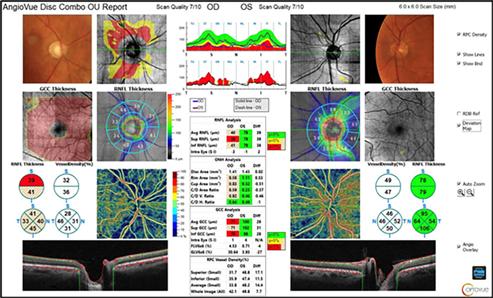
In the case below, a 62-year-old male presented with advanced/severe POAG (based on visual field criteria). The OCT shows severe retinal nerve fiber layer (RNFL) and ganglion cell damage OD. The radial peripapillary capillary (RPC) is also thin.
These issues may be a combination of glaucoma and an old ischemic optic neuropathy, especially considering the overall thinning of the RNFL, and the mild pallor of the neuroretinal rim that was also noted in the right eye. This report highlights a few things:
- Measurements like these help to stage the severity of structural defects at a glance, allowing providers to review inter- and intra-eye symmetry and spend less time reviewing multiple reports to make an informed assessment while spending more time with patients.
- The outer columns for each eye show the deviation map with Pixel x Pixel analysis where the thickness has between a 5% yellow and 1% red chance of being normal. The thickness map shows the loss of the superior and inferior bundle. The RNFL numbers from the 100μ wide ring at 3.45 show
corresponding loss, a larger C/D ratio and significant vessel density loss in both the superior and inferior hemispheres. - The center column gives a tabular format of the results and provides detailed inter- and intra-eye comparison values for the parameters.
 This comprehensive report not only lays out complex data for the physician in a readable manner but also enables me to show my patient his left eye and say, “This is fairly normal and what it should look like, and here is the damage we are seeing in your right eye.” These capabilities are fundamentally impactful to patients understanding their diagnosis and may increase their compliance with treatment and follow up recommendations.

Nate Lighthizer, OD, FAAO
Born and raised in Bismarck, ND, Nate Lighthizer, OD, FAAO, is a graduate of Pacific University College of Optometry. Upon graduation, he completed a residency in Family Practice Optometry with an emphasis in Ocular Disease through Northeastern State University Oklahoma College of Optometry. Dr. Lighthizer has since joined the faculty at the Oklahoma College of Optometry and serves as the Chief of Specialty Care Clinics and the Chief of Electrodiagnostics Clinic. In 2014, he founded and now heads the Dry Eye Clinic at the College of Optometry. Also in 2014, he was named the Director of Continuing Education as well as the Assistant Dean for Clinical Care Services at the Oklahoma College of Optometry. He is a founding member, and currently serves as Vice President, of the Intrepid Eye Society which is a group of emerging thought leaders in optometry. He was named a member of PCON 250—a list of the top 250 optometrists in the country who practice progressively, provide innovative patient care, conduct optometric research or excel in academia and share what they have learned with other optometrists to advance the profession. Dr. Lighthizer lectures nationally on numerous topics, most notably advanced ophthalmic procedures, electrodiagnostics and ocular disease.
