[OCT Article] Multimodal OCT Imaging of Adenovirus Keratitis by Adil El Maftouhi
![[OCT Article] Multimodal OCT Imaging of Adenovirus Keratitis by Adil El Maftouhi Image](https://blog.visionix.com/hs-fs/hubfs/fig1%20-%20adenovirusv2.png?width=1250&name=fig1%20-%20adenovirusv2.png)
CASE HISTORY:
- A 28-year-old woman presenting with persistent bilateral conjunctivitis transmitted by her 2-year-old daughter.
Clinical results:
-
At the biomicroscopic examination, the patient presents bilateral conjunctival hyperemia with nummular lesions involving the anterior stroma in both eyes, accompanied by some photophobia and itching.
-
Corneal OCT reveals subepithelial lesions affecting the basement membrane and anterior stroma in both eyes on the B-scan images (Fig. 1).
-
These subepithelial lesions are clearly highlighted by the En Face OCT (8x8mm) of the Optovue Solix (Visionix) (Fig. 2). Although these lesions may persist over time, they typically resolve without any scarring. En face OCT provides a quantification of these lesions to observe the response to treatment.
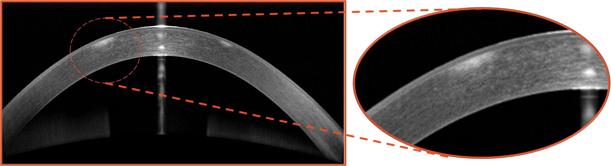
⬆️ Fig 1: 10mm Bscan, Optovue Solix, Visionix. Focal and punctate hyper-reflectivity involving the epithelium and anterior stroma.
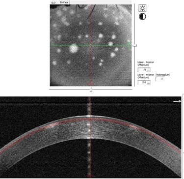 ⬅️ Fig 2: "En Face" OCT 8x8mm with a 30-micron slab positioned between the basement membrane and the anterior stroma.
The nummular lesions are highlighted.
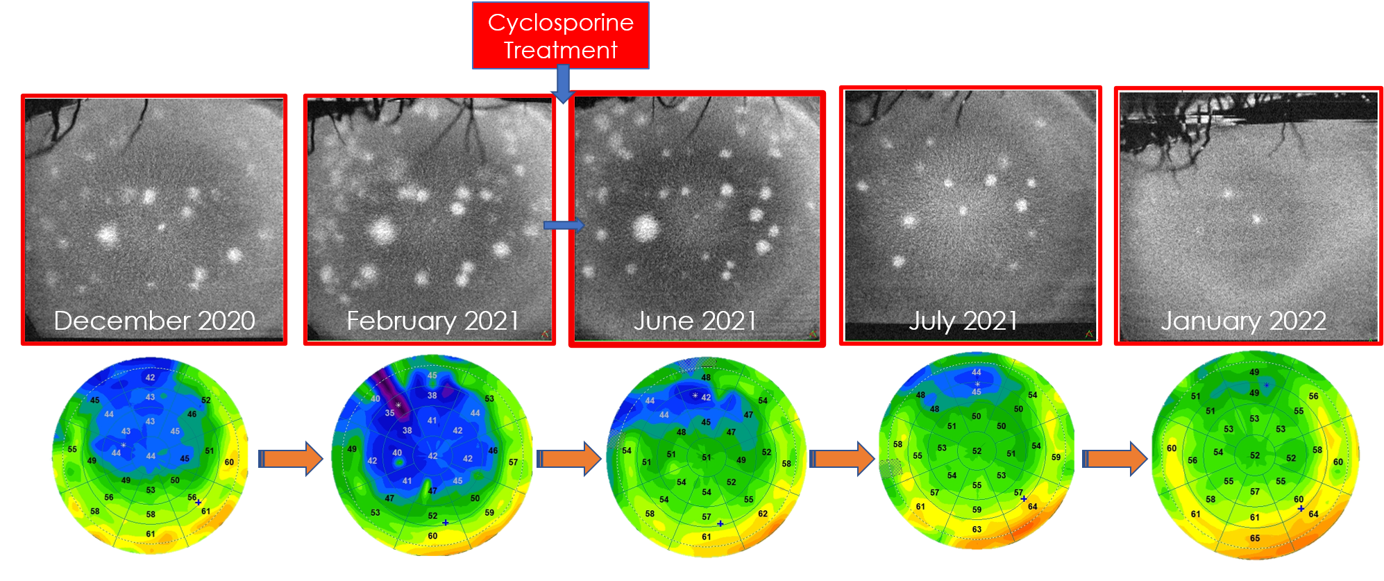 ⬆️ Fig 3: AS EN FACE OCT, OPTOVUE SOLIX, VISIONIX. Nummular Keratitis with anterior stromal infiltrates.
- After initiating Cyclosporine treatment (0.1%), an improvement in the ocular surface is observed, with a reduction in the atrophy of the corneal epithelium. En face OCT demonstrates a decrease in the number of nodular lesions, indicating a positive response to the treatment. Fig 3.
- Twelve months later, there is nearly complete resolution of subepithelial nodules with improvement in the epimapping profile, which normalizes. Fig 3.
Conclusion
- En Face OCT combined with epithelial mapping allows for quantification and monitoring of the progression of Adenovirus Keratitis, providing detailed insights into the treatment response.
- The multimodality of OCT, including B-scan, En Face OCT, and epithelial pachymetry, enables a precise assessment of corneal and ocular surface damage for improved monitoring.


Adil El Maftouhi is an orthoptist specializing in ocular imaging and exploration at the CHNO des XV-XX (Paris) and the Center Ophtalmologique de Rive Geneva in Switzerland. He is the author of international works on OCT and OCT-A. He has also published several peer-reviewed articles such as 'OCT and Dry Eye' and 'OCT: The Intelligence of the Epithelium.'
Adil El Maftouhi is actively involved in the development and utilization of various imaging systems in ophthalmology, leveraging the potential of each system for clinical benefit. He concurrently contributes to the development of new imaging applications and software, particularly in the field of OCT technology. Mr. El Maftouhi is also engaged in clinical and pharmacological research projects in the areas of medical retina, glaucoma, and visual impairment.
The information is intended for general informational purposes only. It is not intended as, and should not be considered, a substitute for professional medical advice, diagnosis, or treatment.
The content is not designed to replace the relationship that exists between a patient and their healthcare provider. Any medical decisions should be made in consultation with a qualified healthcare professional who can provide information tailored to your individual circumstances.
Medical procedures, case studies, and practices mentioned in this content may vary based on regional standards, local regulations, and the discretion of the providing healthcare professional. What may be considered appropriate and ethical in one country may differ in another.
The content may include general references to medical practices, medications, or treatments that are widely accepted in certain regions but may not be applicable or endorsed universally. It is important to consult with a healthcare professional in your jurisdiction to ensure the information is relevant to your specific situation.
The authors, publishers, and contributors of this content disclaim any liability for any adverse effects resulting directly or indirectly from information contained in this content. Readers should exercise their own judgment and seek the advice of healthcare professionals as appropriate.
By accessing and using this content, you acknowledge and agree to the terms of this disclaimer.
