
The True-Color Advantage
In his busy regional eye center, Crockett Eye Clinic in Crockett, Texas, John A. McCall Jr., OD, has built a practice around a simple idea: the right technology should make it easier to find disease, document it clearly and move patients through the clinic without wasting time.
The Ultra-Widefield TrueColor Confocal EIDON is a key tool in the clinical workflow. Dr. McCall says he has long invested in imaging equipment, including three OCT devices, because the clinic functions as a regional referral hub in a medically underserved part of the state. That makes diagnostic precision especially important.
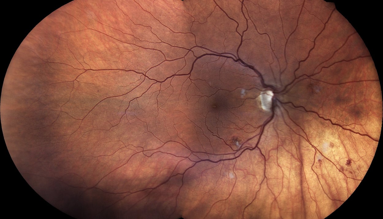
What drew him to the EIDON was its confocal scanning laser technology and its true-color imaging. “Some retinal imaging will give you three colors, like red, green and blue, and simulate what the real color would be, which is often close. But there’s nothing as close as a true-color camera,” Dr. McCall says.
That matters to him because of his reliance on retinal images, especially as his practice grows. Dr. McCall’s son Matthew McCall, OD, and fellow 2026 graduate of Kentucky College of Optometry, University of Pikeville, Carolyn Ogle, OD, have joined the practice along with associate William Pope, OD. The images captured on the EIDON have become a key part of clinical records and legal documents and, often, are the difference between a vague description and a clear diagnosis.
Directed Capture
Dr. McCall recalls the first time he used the device on a small retinal tear located temporally and just below the midline. He asked his tech to show him how the directional capabilities work on the EIDON. “She tapped on the general area where I thought the tear was, and that moved the fixation, bringing that lesion right in front of the camera. She hit a button, and it captured in full color.”
That robotic targeting is one of the system’s biggest advantages, he says. Instead of relying on a broad, one-shot image or overlapping images and hoping the lesion appears, the EIDON lets the user direct the camera to a specific zone of interest. If a pathology is in the periphery, the operator can bring it into view, center it and capture it quickly.
“It’s very quick, very easy,” Dr. McCall says. The operator can touch the area on the screen, shift fixation and bring the pathology into the center of the image. That helps the team capture what they need without a complicated setup or repeated imaging.
“A true color image gives you a really, really good basis for following these patients.” - Dr. McCall
Cleaner Workflow
Dr. McCall says the EIDON also fits into the practice’s workflow in a deliberate way. His team uses it in a diagnosis-driven model, not as a routine screening tool for every patient. “We don’t want to use the same device for screening and pathology,” he says. The clinic stores screening images and diagnostic images in separate locations, which helps maintain a cleaner process for coding and billing and clearer documentation.
He says the practice has structured imaging around the idea that different instruments serve different purposes. The EIDON is used when the question is diagnostic and documentation-focused, while other devices handle screening or broader imaging tasks. That distinction helps the clinic maintain compliance while maximizing efficiency.
Dr. McCall says the technology also improves patient flow. In a practice where technicians complete the imaging before the doctor sees the patient, anything that captures quickly and accurately reduces bottlenecks. And for older patients in a rural area, fewer steps and less moving around between devices can make the experience easier.
Plus, the EIDON’s confocal design also helps with image quality, he says. Unlike some flash-based systems that can constrict the pupil between exposures, he says the confocal approach allows multiple clean images without any compromise to image quality. That helps preserve consistency while reducing the need for repeated attempts. “I like to have good clean images,” Dr. McCall says. “What you see is what you get with this device, and it’s true color.”
That is also helpful when working with patients whose conditions require long-term comparison, such as glaucoma and macular degeneration. In glaucoma care, he wants a clear baseline image of the optic nerve and cupping. In retinal disease, he wants documentation that is easy to review over time.
The quality of the images also aids documentation and patient education. He—or whichever doctor sees the patient on the next visit—doesn’t have to rely on a description or drawing. “A true color image of that gives you a really, really good basis for following these patients,” Dr. McCall says.
Dr. McCall’s father opened the practice in 1950; and an advanced level of care is part of its larger legacy of service. He jokes with associates that this office is so well-equipped with advanced technology that they are now spoiled for anyplace else. But that’s exactly how he wants it. He says that tools like the EIDON help him deliver both the clinical rigor and the efficiency his patients need.
Standout Features of the iCare EIDON Widefield TrueColor Confocal Fundus Imaging System
.png?width=235&height=233&name=iCare_EIDON_with-screen-and-logo%20(2).png)
- Multiple imaging modalities including TrueColor, blue, red and Red-Free and infrared confocal images
Widefield, ultra-high-resolution imaging Capability to image through cataract and media opacities Dilation-free operation (minimum pupil 2.5 mm) Flexibility of fully automated and fully manual mode All-in-one compact design, no additional PC required
About the Author

JOHN A. McCALL JR., OD
Dr. John A. McCall, Jr. is a distinguished optometrist and leader in the field of eye care. A 1977 graduate of the University of Houston College of Optometry, he has dedicated his career to advancing optometric care, clinical education, and patient outcomes. He is a former President of both the Texas Optometric Association and the American Optometric Association and is Co-Founder and current President of the American Academy of Optometric Medicine & Surgery.
Dr. McCall is a pioneer in glaucoma treatment and holds the patent for the Rapid SLT Lens, one of the leading glaucoma surgery lenses worldwide. He has more than a decade of surgical experience and has performed procedures at his surgery center in Logansport, Louisiana. His contributions to optometry have earned him numerous honors, including Texas Optometrist of the Year, the Texas Distinguished Service Award, and University of Houston College of Optometry Alumni of the Year.
As Co-Founder and Senior Vice President of Vision Source, Dr. McCall has also played a significant role in advancing the practice of optometry and supporting independent optometric professionals. He continues to practice optometry in Crockett, Texas.
**The views and experiences expressed are those of the individual user. They may involve off-label use of the medical device, which is not endorsed or approved by the manufacturer. Medical procedures, case studies, and practices mentioned in this content may vary depending on regional standards, local regulations, and the discretion of the healthcare provider. Not all products, accessories, services, or offers are approved in every market.
This article originally appeared in the September 2026 issue of Vision Source OD magazine - Vision Source OD • 3QTR 2026.