Advanced Imaging Improves Decisions and Patient Outcomes

Eye Surgeons of Indiana practice has grown tremendously over the years since I came onboard in 2002. At present, we have 20 doctors covering seven locations and three surgical centers. As a partner in the practice for the past six years, I’ve been able to help build our surgical volume by more than 200%.
In our growing and evolving business, I’m always looking out for smart opportunities to incorporate new instrumentation. Our goal with new technology is often focused on enhancing patient care and improving efficiency.
Expectations Exceeded
One of our recent technology acquisitions was the Optovue Solix by Visionix, the next generation FullRange® OCT and OCT-Angiography with a multitude of imaging options, enhanced metrics, integrated fundus camera, and external IR imaging. We added this instrumentation about a year ago, following other positive experiences with Optovue devices throughout all of our locations.
I envisioned that the Optovue Solix OCT/OCT-A would enhance our care experience, providing more thorough data for patients, as well as an efficient experience for patients and staff. It also added some options we didn’t have before with our OCT technology, including topography, anterior segment images, and the latest OCT-A technology. The advances in analytics from the data allow us to work smarter.
This technology exceeded my own and my colleagues’ expectations. It allows us to capture information that we would not be able to otherwise. One feature that stands out is the anterior segment imaging with the ability to get quick images from the anterior and posterior—top to bottom and front to back. These corneal scans include pachymetry, angle measurements, and 3D images.
Easy Implementation in Many Situations
The Optovue Solix OCT/OCT-A is making an impact from the screening and wellness scans to tracking patients with ocular disease and their progression over time. Our practice provides 100 percent medical eye care, so we are often using this technology for medical indications that are then billed to insurance. However, we still do use the screening exam function as part of our standard workup for cataract patients. That scan is built into our exam fee.
The opportunity is right in front of us since every single patient who comes to our offices has a medical condition of the eye. Here are a few examples of how this technology is put to work in our offices every day.
-
Screening exam: Look for changes that are consistent with certain conditions such as early glaucoma. Rule out macular pathology in patients pursuing refractive cataract surgery.
-
Glaucoma: Analyze if we need to do a further workup with technology that produces a 3D scan for precise measurement of nerve fiber and change over time. Identify patients that are progressing and may benefit from interventional glaucoma treatment including SLT, MIGS, and drug delivery devices.
-
Retina: Monitor if treatment is working, if we need to do injections, and progression of disease with the new high-density 3D scans. Identify non-exudative choroidal neovascularization in AMD patients that may require closer monitoring.
-
Keratoconus: Use anterior segment capabilities to assess and monitor patients, as well as track our patients undergoing crosslinking.
-
Diabetes/retinal vascular conditions/macular degeneration: Use OCT-angiography for better management of patients with these conditions.
Efficiency and Ease of Use
Imaging with the Optovue Solix OCT/OCT-A helps simplify the exam process for patients. It’s more efficient because it allows us to monitor patients without using multiple devices. For retina cases, we don’t need to take as many photographs as we used to in order to see what we need for patients with age-related macular degeneration, and in particular, for those with geographic atrophy.
Training was excellent, and the support has been, too. If we ever have a question or need to troubleshoot, the Visionix support team is there to help us.
Imagery for Evaluation and Education
Personally, I share images with patients selectively. When I review, I’m making sure that I have the best information to make a good decision for each patient. For me, it’s the data and nuances that can be revealed that is the true value in this instrumentation.
However, also in our practice, our medical retinal specialist has a practice style where he shares images more often. There are benefits to showing the images to patients to validate decisions or celebrate when they have a treatment success.
I also use the images to share with referring outside providers. Many of those colleagues don’t have oct-angiography or anterior segment imaging. The scans are also an asset for educational purposes when I’m participating in CE events.
Enhanced Decision Making
Recently, we used our Optovue Solix OCT/OCT-A with a patient who came in with persistent corneal edema after cataract surgery. The clinical exam was challenging, and the source could not be determined.
We very easily imaged that patient with the anterior segment mode. It revealed a problem with the layer of the cornea that was easily fixable.
With a minor surgical intervention, it was successful at addressing the complication. The Optovue Solix OCT/OCT-A put the data behind the decision to see if we needed to go back to surgery or not.
We’ve also had multiple patients where we have seen progression of disease, such as glaucoma, in their OCT images. From a retina standpoint, I’ve detected very subtle changes in patients’ high-risk macular degeneration. This allows us to diagnose early and be able to prevent vision loss, thanks to OCT-angiography.
This technology helps confirm our recommendation to escalate their therapy or move to surgical intervention or implantation of a medication device. We can make smarter decisions and intervene appropriately.
OPTOVUE SOLIX OCT/OCT-A BY VISIONIX
- The ONLY OCT with FDA-cleared OCT-A metrics
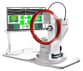
- FullRange® Retinal 16x6.25mm scan
- FullRange® Anterior Chamber 18x6.25mm scan
- Ultra-fast 120kHz scan speed
- Higher scan density & precision vs. other OCTs/OCT-As
- Integrated fundus camera
- External color & IR imaging
- New optional Topography Module available!

Damon dierker, od, faao
Damon Dierker, OD, FAAO, is the Director of Optometric Services at Eye Surgeons of Indiana and an adjunct faculty member at the Indiana University School of Optometry. He practices consultative optometry with a special interest in ocular surface disease, glaucoma, retina, and peri-operative care. Dr. Dierker has developed a dedicated dry eye clinic within his practice and helps colleagues across the country in this area as the creator of Dry Eye Boot Camp and co-founder of Eyes on Dry Eye. Additionally, he is Past President of the Indiana Optometric Association. To contact him: damon.dierker@esi-in.com
This article originally appeared in Review of Optometric Business in February 2026: https://reviewob.com/advanced-imaging-improves-decisions-and-patient-outcomes/
**Medical procedures, case studies, and practices mentioned in this content may vary based on regional standards, local regulations, and the discretion of providing healthcare professional. What may be considered appropriate and ethical in one country may differ in another.
