Abstract
Keratoconus is a bilateral, asymmetric, noninflammatory disease characterized by progressive corneal thinning resulting in corneal protrusion and irregular astigmatism. It affects both sexes and all ethnicities and results in distorted and decreased vision. Onset often occurs at puberty and stabilizes at midlife, and although early and late-stage cases can occur, they are uncommon. The incidence of keratoconus in the general population was reported to be 54.5 per 100,000 in 1986, 1 per 2,000 in 1998, and 1 per 375 in 2017. This tremendous increase in reported prevalence may be the result of advanced diagnostic tools being used for early detection throughout the past two decades. Historically, the diagnosis of keratoconus was based on subjective symptoms and clinically significant objective findings that were able to be observed on physical examination. Although keratoconus does have well-described clinical signs, early forms of the disease often go undetected, with a mean age at diagnosis reported as 28 years. The classical management for keratoconus has long been based on improving visual acuity with contact lenses, specifically rigid gas permeable lenses, and monitoring the disease. However, nothing could be done to stop the progression of the disease. Over time, approximately 10% to 20% of patients with keratoconus needed a penetrating keratoplasty due to factors, such as corneal scarring, steeper corneal curvature, worse visual acuity, contact lens discomfort, and poorer vision-related quality of life.
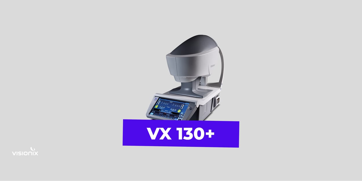
The VX 130+ is a multifunctional diagnostic tool that can be used in any practice setting. Although it is a small machine, the VX 130+ combines seven tools that provide improved diagnostic efficiency, increased documentation, automated calculations, and consistent measurements for not just general needs but all relevant in the diagnosis of keratoconus. Due to the multifunctionality, a multitude of test results are provided, including mesopic and photopic refraction, topography, pachymetry, and tonometry. The VX 130+ provides a complete bilateral analysis in less than 2 minutes, including anterior and posterior keratometry, anterior and posterior topography, mesopic and scotopic refraction, anterior chamber depth, corneal thickness, eccentricity, noncontact and compensated tonometry, corneal asphericity, pupillometry, and ocular aberrations. The VX 130+ provides a tremendous amount of relevant patient data, is cost-effective, time efficient, and can be used on all patients in any clinical office.

