Case Study: Multiple Imaging Techniques to Detect Corneal Irregularities
A comprehensive corneal assessment is fundamental before both refractive and cataract surgery. Detecting corneal irregularities before surgery using a combination of corneal topography or tomography in addition to epithelial mapping is essential. Using both of these technologies drives decisions with regards to performing corneal cross-linking, selecting the appropriate type of refractive surgery, and selecting the intraocular lenses that provide the patient with their desired outcome.
Placido disk technology is an example of corneal topography which provides a 2D map showing variations in corneal curvature. A placido disk topographer projects concentric rings onto the eye to evaluate the cornea shape. Placido disk topographers depend on a stable tear film, therefore patients with dry eyes may be challenging to image. Furthermore, this technology only focuses on the anterior cornea and is unable to provide posterior cornea structure information. Posterior corneal elevation changes can precede anterior changes, therefore corneal tomography which provides a three-dimensional assessment of the cornea may be a superior option. Scheimpflug photography and Optical Coherence Tomography (OCT) are examples of corneal tomography technologies that generate curvature, power, elevation and pachymetry maps. The added benefit of Optical Coherence Tomography is epithelial mapping as it can detect anterior stromal change through epithelial thinning. This occurs prior to any change in the surface curvature which is needed for detection by Placido or Sheimpflug.
In this study, images from three different imaging modalities were acquired on 28 eyes of 14 patients. Subjects were imaged on Zeiss Atlas with Placido technology, Oculus Pentacam with Scheimpflug technology, and Optovue Solix with OCT technology. The first image for each device was compared. The population included: one normal cornea, two contact lens wearers, one dry eye, five keratoconus, three irregular steepening, one post-LASIK, and one flat cornea.
The cornea elevation maps were compared between the three devices and visually demonstrated comparable presentations. The values compared between the three devices were: K1 and K2. K1 and K2 represent the dioptric power of the flattest and steepest meridians of the cornea and can help in diagnosing keratoconus and ectasia. Overall, values were found to be comparable between the three devices for K1. For K2, Optovue Solix and Pentacam were comparable (p=0.07), however both devices were statistically different from Atlas (p=0.00029, p=0.0314). The outliers were all patients with moderate to severe keratoconus. Though statistically different, the clinical interpretation of K2 between the three devices was comparable.
Additionally, pachymetry apex was compared between the Pentacam and Optovue Solix and showed that the Optovue Solix pachymetry apex was on average 20 microns thinner than Pentacam. The difference may be attributed to differences in axial resolution between the two devices. OCT has a 5-micron axial resolution while Scheimpflug has a 15-micron axial resolution.
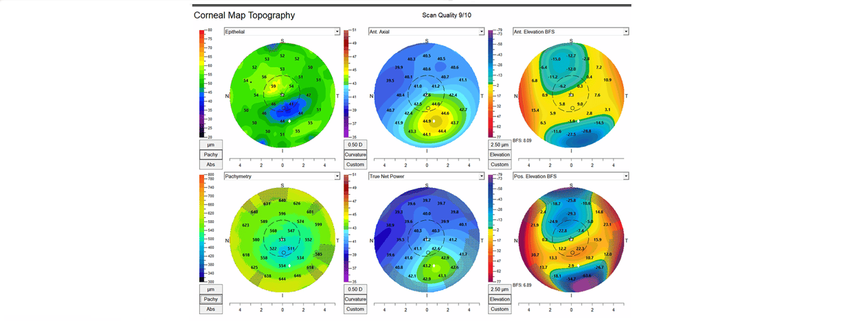
Overall, the Optovue Solix is able to demonstrate both epithelial mapping in addition to tomography, thereby providing a doctor with comprehensive data to help plan for a favorable surgical outcome. While corneal topography and tomography can demonstrate keratoconus, forme fruste keratoconus can be challenging to distinguish without the use of epithelial mapping. Steepening of the cornea with associated thinning of the epithelium is consistent with forme frustre keratoconus and may prompt a corneal cross-linking procedure. If a patient with such epithelium presentation is interested in refractive surgery, an ICL procedure with topography-guided PRK touch-up may be suggested. The image below demonstrates the epithelial map with pachymetry on the far-left side next to the associated tomography maps for axial, true net power anterior elevation, and posterior elevation maps. In addition to epithelial mapping, posterior elevation maps can also show early ectasia.
In conclusion, combined imaging modalities can be used to detect early corneal irregularities. Optovue Solix provides anterior elevation maps and posterior elevation maps like other devices; however, the added epithelial thickness map is specifically useful in early keratoconus detection.

Figure 1. Corneal Map Topography from the Optovue Solix showing the epithelial map with pachymetry on the far-left side next to the associated tomography maps for axial, true net power, anterior elevation, and posterior elevation maps.
 Figure 2. Corneal Map Topography from the Optovue Solix showing anterior and posterior tangential, mean curvature, and refractive maps.
OPTOVUE SOLIX BY VISIONIX
- The ONLY OCT with FDA-cleared OCT-A metrics
 FullRange® Retinal 16x6.25mm scan
- FullRange® Anterior Chamber 18x6.25mm scan
- Ultra-fast 120kHz scan speed
- Higher scan density & precision vs. other OCTs/OCT-As
- Integrated fundus camera
- External color & IR imaging
- New optional Topography Module available!
See the Optovue Solix live at the American Academy of Optometry Booth #702 and the American Academy of Ophthalmology Booth #1441.
 Dr. Ella Faktorovich is the Founder and Director of Pacific Vision Institute. Under her leadership, Pacific Vision Institute is often the first in the Bay Area to bring the industry's newest diagnostic and treatment technologies to patients. Dr. Faktorovich has been listed in the Best Doctors in America and in the Top 100 Health Professionals in the World. She has been selected by Castle Connolly's Top Doctor for more than 15 years in a row as one of the best ophthalmologists in San Francisco Bay Area. Dr. Faktorovich has also been chosen as a Top Doctor in San Francisco to be included in the annual Best Doctors issue of San Francisco Magazine. Dr. Faktorovich is the founder and remains the Chair of the Annual Advances in Eye Care San Francisco Symposium dedicated to continuing education of the Bay Area eye doctors in refractive surgery and other aspects of advanced patient eye care.
 Dr. Katherine Makedonsky is an associate optometrist at Marina Village Optometry. She focuses mainly on clinical research but continues with patient care on a part-time basis. She is the Clinical Director at Visionix and collaborated with Dr. Faktorovich on this research project.
**The information provided is for general informational purposes only. It is not intended to replace and should not be considered a substitute for professional medical advice, diagnosis, or treatment. The content is not designed to replace the relationship between a patient and their healthcare provider. Any medical decision should be made in consultation with a qualified healthcare professional who can provide information tailored to your individual situation. Medical procedures, case studies, and practices mentioned in this content may vary depending on regional standards, local regulations, and the discretion of the healthcare provider. The views and experiences expressed are those of the individual user. They may involve off-label use of the medical device, which is not endorsed or approved by the manufacturer. What may be considered appropriate and ethical in one country may differ in another. The content may include general references to medical practices, medications, or treatments that are widely accepted in certain regions but may not be universally applicable or approved. It is important to consult a healthcare professional in your jurisdiction to ensure that the information is relevant to your specific situation. The authors, editors, and contributors of this content disclaim any liability for any adverse effects resulting directly or indirectly from the information contained herein. Readers should exercise their own judgment and seek advice from healthcare professionals when necessary. By accessing and using this content, you acknowledge and accept the terms of this disclaimer.
