

Case Study: Detection of Diabetic Macular Edema Despite Minimal Symptoms
Introduction
Diabetic retinopathy remains one of the leading causes of vision loss among adults, yet vision threatening diabetic retinal disease may develop even in patients who report no visual complaints and have little or no clinically apparent diabetic retinopathy.1-3 This case highlights the importance of careful comparison of serial retinal imaging and the value of optical coherence tomography (OCT) in identifying diabetic macular edema (DME) in the presence of an epiretinal membrane (ERM) that was not readily apparent during the clinical examination.
Case Report
A 77-year-old male presented for his annual comprehensive eye examination. His ocular history was significant for uncomplicated cataract surgery performed three years earlier. His medical history was notable for diabetes, hypertension, and hypercholesteremia for which he was being medically managed. He had no previous history of diabetic retinopathy.
The patient reported no subjective change in his vision since his previous examination. Entering visual acuity was OD 20/50, OS 20/50-1. His best corrected visual acuity measured OD 20/40 and OS 20/50. Examination of the anterior segment was unremarkable, with a well-positioned posterior chamber intraocular lens in each eye.
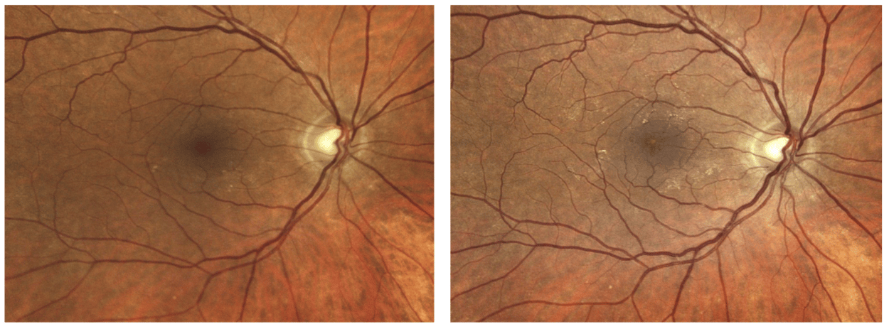
Widefield fundus photography (iCare EIDON) revealed a subtle, irregular yellow lesion within the macular region. Closer inspection demonstrated a small ring-like abnormality located within the macula. (Figure 1) Although the retinal findings appeared relatively subtle during the examination, comparison with fundus photographs obtained the previous year demonstrated a clear change in appearance. (Figure 2 and 3)
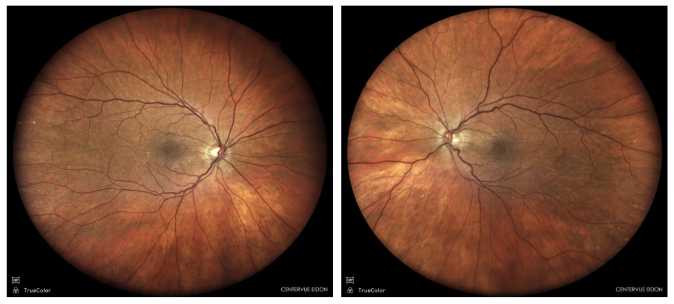 Figure 1. Entering fundus presentation of the patient’s right and left eyes in 2026 acquired with an EIDON imaging system.
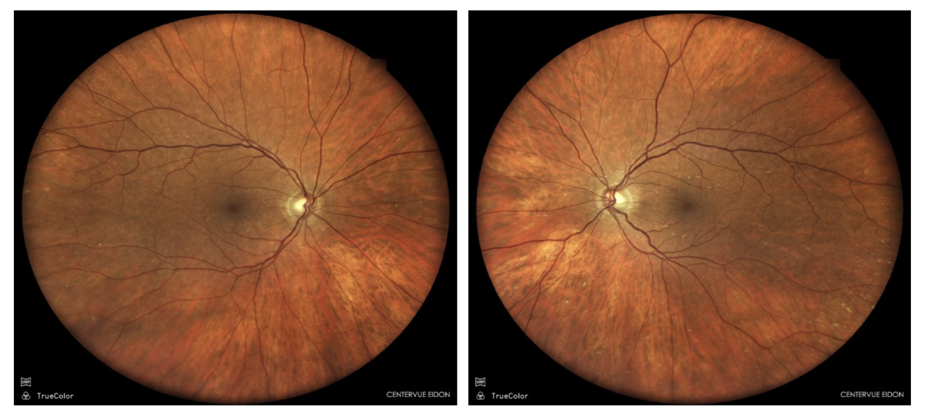 Figure 2. Fundus photographs of the patient’s right and left eyes in 2025 acquired with an EIDON imaging system.
The unexplained reduction in best corrected visual acuity and the newly identified retinal abnormalities warranted a spectral domain OCT imaging of the macula. OCT (Optovue Solix) demonstrated significant bilateral macular edema characterized by increased central retinal thickness (Figure 3) and multiple intraretinal cystic spaces extending through the central macula along with a mild epiretinal membrane. (Figure 4)
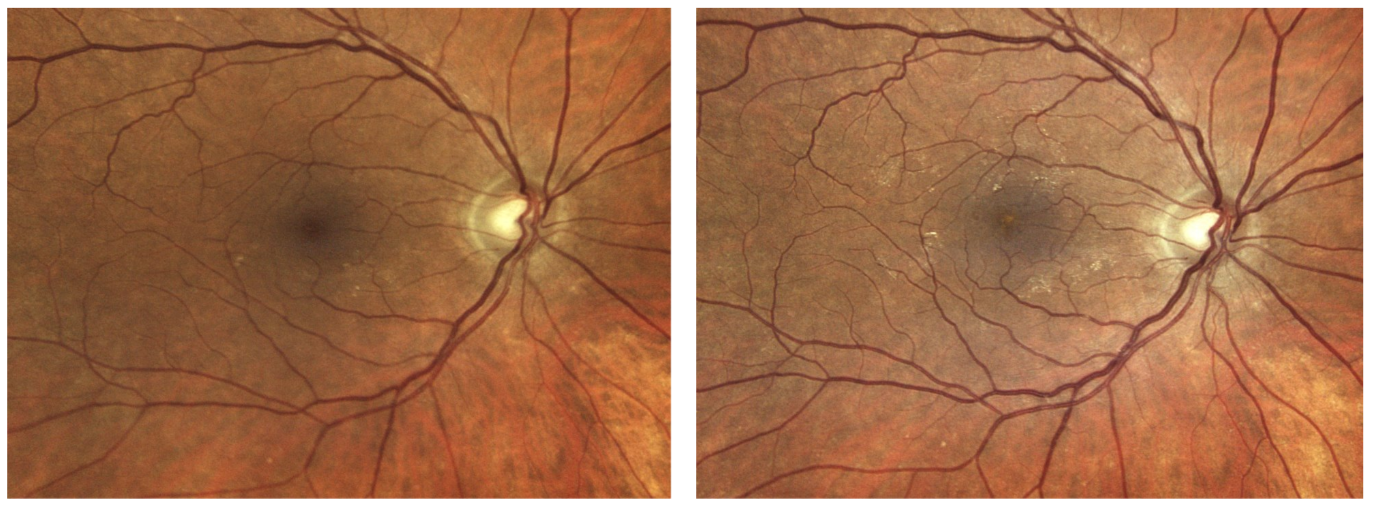 Figure 3. Patient’s right eye in 2025 compared to 2026, acquired with an EIDON imaging system.
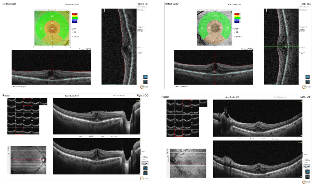 Figure 4. Increased macular thickness confirmed with the Optovue Solix OCT.
The patient was diagnosed with bilateral diabetic macular edema with an epiretinal membrane. This case illustrates that clinically significant diabetic macular edema may develop despite minimal symptoms and relatively subtle funduscopic findings, emphasizing the importance of multimodal retinal imaging in diabetic patients.
The examination findings and OCT images were reviewed with the patient in detail. Given the presence of clinically significant bilateral diabetic macular edema, an immediate referral was made to a retina specialist for further evaluation and management. The patient understood the findings and the importance of timely retinal intervention to preserve vision.
Discussion
This case demonstrates several important clinical lessons. Patients with diabetes may develop visually significant diabetic macular edema while remaining largely asymptomatic. Small changes on fundus photography may represent significant underlying retinal pathology. Comparing current retinal images with previous photographs can reveal subtle progression that may otherwise be overlooked.
The OCT remains the gold standard for confirming the presence, location, and severity of diabetic macular edema and should be obtained whenever retinal abnormalities or unexplained reductions in visual acuity are identified. Early detection and prompt referral to a retina specialist are critical to preserving long-term visual function. This case demonstrates this important concept.
References
- Wong TY, Sun J, Kawasaki R, et al. Guidelines on diabetic eye care: The International Council of Ophthalmology recommendations for screening, follow-up, referral, and treatment based on resource settings. Ophthalmology. 2018;125(10):1608-1622.
- Browning DJ, Stewart MW, Lee C. Diabetic macular edema: Evidence-based management. Indian Journal of Ophthalmology. 2018;66(12):1736-1750.
- Schmidt-Erfurth U, Garcia-Arumi J, Bandello F, et al. Guidelines for the management of diabetic macular edema by the European Society of Retina Specialists (EURETINA). Ophthalmologica. 2017;237(4):185-222.
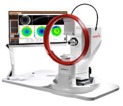
OPTOVUE SOLIX oct/oct-a
- The ONLY OCT with FDA-cleared OCT-A metrics
 FullRange® Retinal 16x6.25mm scan
- FullRange® Anterior Chamber 18x6.25mm scan
- Ultra-fast 120kHz scan speed
- Higher scan density & precision vs. other OCTs/OCT-As
- Integrated fundus camera
- External color & IR imaging
- Optional Topography Module available
icare eidon widefield fundus imaging
- Multiple imaging modalities including TrueColor, blue, red
.png?width=203&height=201&name=iCare_EIDON_with-screen-and-logo%20(2).png) and Red-Free and infrared confocal images
and Red-Free and infrared confocal images Widefield, ultra-high-resolution imaging Capability to image through cataract and media opacities Dilation-free operation (minimum pupil 2.5 mm) Flexibility of fully automated and fully manual mode All-in-one compact design, no additional PC required
This article originally appeared in Optometric Management in September 2026 - https://optometricmanagement.com/issues/2026/september-october/detection-of-diabetic-macular-edema-despite-minimal-symptoms/

Mile Brujic, OD, FAAO is a 2002 graduate of the New England College of Optometry. He is a partner of Premier Vision Group, a successful four location optometric practice in Northwest Ohio. He practices full scope optometry with an emphasis on ocular disease management of the anterior segment and specialty contact lenses. He is active at all levels of organized optometry. Dr. Brujic is on the editorial board for several optometric publications. He has published over 500 articles and has given over 2,400 lectures, both nationally and internationally on contemporary topics in eye care.
**The views and experiences expressed are those of the individual user. They may involve off-label use of the medical device, which is not endorsed or approved by the manufacturer. Medical procedures, case studies, and practices mentioned in this content may vary depending on regional standards, local regulations, and the discretion of the healthcare provider. Not all products, accessories, services, or offers are approved in every market.