OCT-A, invented by Optovue, celebrates a decade of innovation

Optical Coherence Tomography (OCT), a non-invasive imaging technique, developed in 1991, has since been used to visualize the different layers of the retina invisible with a traditional fundus. Twenty-three years later, OCT-A, designed by Professor David Huang's team for Optovue in 2014, emerged as an extension of OCT via the integration of an algorithm called SSADA (Split Spectrum Amplitude Decorrelation Angiography). Optovue's XR Avanti thus enabled the first OCT-A examinations. In 10 years, nearly 2,500 devices equipped with Optovue OCT-A technology have been installed around the world. This anniversary is also an opportunity to pay tribute to Professor Bruno Lumbroso, who recently passed away. As one of the founding pillars of modern macular imaging, he has indeed greatly contributed to the understanding and democratization of OCT-A.
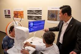
David Huang, M.D., Ph.D., (right) demonstrating the OCT-A technology he co-invented, at Oregon Health & Science University in Portland, Oregon. (courtesy of Mr. David Huang, M.D., Ph.D.)
Unlike OCT, which focuses on the tissue structure of the retina, OCT-A allows arterial, venous and capillary vessels to be visualized in high resolution and in three dimensions. Thanks to OCT-A, clinicians can detect the finest vascular abnormalities early and monitor their development on a regular basis.
This technology has revolutionized the diagnosis and management of numerous pathologies such as diabetic retinopathy, AMD, capillary occlusions and glaucoma. Until now, fluorescein or indocyanine green angiography was the reference method for visualizing retinal and choroidal vascularity. Conversely, OCT-A can be performed without a contrast product, thus eliminating the intravenous, the constraints of a long and rigorous protocol, as well as the adverse effects, the allergic or even anaphylactic risk linked to contrast products. , which, although rare, are real.
“It’s a revolution. The examination lasts a few tens of seconds. OCT-A has changed paradigms whether for diagnosis or patient monitoring,” confirms Doctor Alexandra Miere, MCU-PH at CHI Créteil.
A technology that has been evolving for 10 years
Over the past decade, Optovue's OCT-A has seen significant improvements. Whether in terms of eye movement compensation, image resolutions, artifact reductions, acquisition speed, acquisition window size, increased from 3x3 and 12x12 for the latest Optovue Solix model. The image quality has thus been improved. New biomarkers, such as parafoveal or peripapillary vascular density, have also been added.
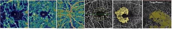
Quantitative analyzes provided by OCT allowing the therapeutic response to be evaluated and the treatment to be influenced (courtesy of Mr. Adil El Maftouhi, Geneva).
Indiscutable Clinical Progress
OCT-A now allows increased precision in semiological analysis, visualization of neovascular architecture, diagnosis and, ultimately, greater responsiveness in the administration of treatments and better evaluation of their effectiveness.
“With OCT-A, we can detect the presence of new vessels even before the first signs of exudation. However, the visual and functional prognosis will be better if the treatments are carried out much earlier,” explains Mr. Adil El Maftouhi, Orthoptist specializing in ocular imaging in Geneva.
The Lariboisière hospital was one of the first centers equipped with OCT-A. Doctor Ali Erginay can therefore also testify to the way in which it has changed his practice.
« L’OCT-A a été une grande révolution. Avec l’angiographie, vous voyiez les vaisseaux en 2D. Avec l’OCT-A, vous voyez les vaisseaux couche par couche, en 3D. C’est devenu une technique indispensable qui a modifié certaines conduites à tenir, certains schémas thérapeutiques et les fréquences de suivi. Grâce à ce système, la classification de la DMLA a été modifiée. L’OCT-A est devenu un examen de première intention dans plusieurs domaines. Aujourd’hui, elle anime les débats. Va-t-elle, à termes, remplacer l’angiographie classique et l’ICG ? En fait, pour certaines pathologies, c’est déjà le cas », observe-t-il, enthousiaste.
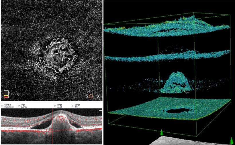
3D OCT-A visualization in a case of Best's disease to better identify the morphology of new vessels (courtesy of Mr. Adil El Maftouhi, Geneva).
Dynamic research and promising prospects
OCT-A, because it makes it possible to collect a significant amount of information on different biomarkers, and on a frequent basis, was quickly adopted by researchers. The number of scientific articles devoted to vascular pathologies, following studies carried out with OCT-A, have increased dramatically over the past 10 years. There are several thousand of them around the world.
OCT-A has thus become a valuable tool in the approach to retinal diseases. Especially since software improvements have been made to facilitate the interpretation of images and to automate the quantitative analysis of the data. In this regard, artificial intelligence, combined with OCT-A, should play a central role in the years to come to facilitate the early detection of pathologies such as choroidal neovascularization.
“OCT-A continues to amaze us” assures Doctor Ali Erginay.
